Test Complete
- Questions
- Score
- Minutes
| Overall Results | |
|---|---|
| Total Questions |
| Category Results | |
|---|---|
Sinus Rhythms
Category: Cardiology
Topic: 3 Lead Rhythms originating in the Sinus Node and Atria
Level: Paramedic
Next Unit: Premature Atrial Complexes
17 minute read
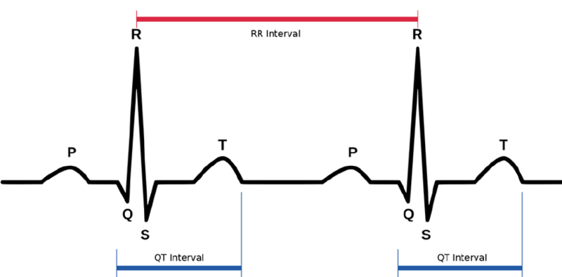
REVIEW: ECG ("EKG") Waveforms and Complexes
The ECG interprets cardiac rhythm, the conduction system, and can even detect myocardial ischemia. The ECG can detect other abnormalities such as valvular heart disease, cardiomyopathy, pericarditis, and hypertensive disease. It is also useful in the followup for drug treatment, e.g., for arrhythmias.
The electrocardiogram (ECG) is a plot of voltage on a vertical axis against time on a horizontal axis.
P-QRS-T
ECG waves are labeled alphabetically starting with the P wave, followed by the QRS complex and then the ST-T complex (ST segment and T wave). A "J point" is the junction between the end of the QRS and the beginning of the ST segment.
The PR interval is measured from the beginning of the P wave to the first part of the QRS complex.
The QT interval consists of the QRS complex which represents only a brief part of the interval, and the ST segment and T wave which are of longer duration.
Normal Sinus Rhythm—"NSR" (60-100 BPM)
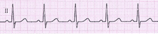
Sinus rhythm is any cardiac rhythm where depolarization of the cardiac muscle begins at the sinus node, characterized on an ECG by the presence of a correctly oriented P wave, and has a rate of 60-100 beats per minute.
Normal conduction indicates that the myocardium is not irritable or injured. The real test to determine whether a patient is hemodynamically stable is to check his or her blood pressure.
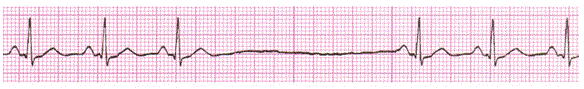
Sinus Bradycardia (< 60 BPM)
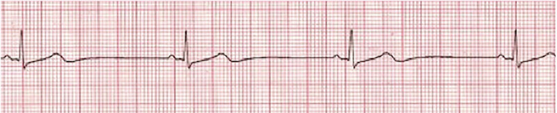
Sinus bradycardia is a rhythm in which the rate of impulses originating in the SA node is lower than normal--a rate of 60 beats per minute or less.
Very few patients, however, will actually experience symptoms unless their heart rate drops to < 50 beats per minute, perhaps the mid 40's. The action potential responsible for this rhythm originates in the sinus node, causing a P wave on the surface ECG tracing that's normal in amplitude and vector. The P waves will typically be followed by a normal QRS complex and T wave.
This rate is a result of a complex interaction between the sympathetic and parasympathetic nervous systems. It also varies by age and physical condition. It can also vary with a sinus arrhythmia due to respiration effects on the SA node.
- Normal Sinus Bradycardia: A normal sinus rhythm has a normal P wave vector on ECG and the rate is largely regular, whereas sinus bradycardia is the same but with a lower rate (<60).
Sinus bradycardia is normal in endurance athletes due to their increased stroke volume, requiring less of a heart rate to adequately oxygenate tissues.
- Abnormal Sinus Bradycardia:
- Sick Sinus Syndrome (SSS). SSS is a dysfunction of the SA node due to age, causing sluggish or absent pacemaking impulses.
Symptoms are fatigue, lightheadedness, palpitations, and syncope. ECG abnormalities include inappropriate heart rate response to activity, sinus pause, and sinus arrest, often without escape beats.
- Sick Sinus Syndrome (SSS). SSS is a dysfunction of the SA node due to age, causing sluggish or absent pacemaking impulses.
POSSIBLE CAUSES:
- Medications. Side effects and toxicity due to parasympathomimetic agents (acetylcholine), sympathetic blockers (beta blockers), opioids and sedatives, cimetidine, digitalis, calcium channel blockers (verapamil), hepatitis C drugs, lithium, and chemotherapy.
- Acute MI.
- Obstructive Sleep Apnea.
- Vagal Stimulation. Heightened parasympathetic activity + sympathetic withdrawal on the SA node: carotid sinus stimulation, vomiting, coughing, Valsalva maneuver.
- Increased intracranial pressure.
- Infection.
Symptomatic bradycardia exists when the following 3 criteria are present:
- The heart rate is slow.
- The patient has symptoms of hypoperfusion.
- The symptoms are suspected of being caused by the slow heart rate.
ECG in Bradycardia of Sinus Origin
A bradycardia of sinus origin will display the following on ECG:
Upright P wave, leads I, II, and aVL
AND
Negative P wave in lead aVR
Treatment of Symptomatic Sinus Bradycardia:
Mnemonic for treatment of symptomatic bradycardia, PACE em! Pacing Always Ends Danger ("P.A.E.D."):
- transcutaneous Pacing,
- Atropine,
- Epinephrine, and
- Dopamine!
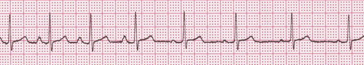
Sinus Tachycardia (> 100 bpm)
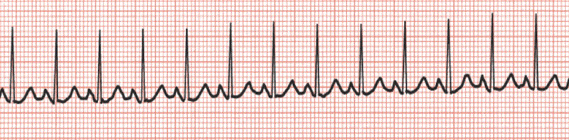
Sinus tachycardia is a rhythm in which the rate of impulses originating in the SA node is elevated from the normal--exceeding 100 beats per minute or more. The impulses originate in the SA node, but it is pacing the heart at a faster rate than normal.
CHARACTERISTICS
- Regular, normal width, P waves present, regular, upright,
- 1 QRS for every P wave,
- R-R Rate > 100, in excess of what one would expect with associated exertion,
- normal PR interval.
CAUSES
- Fever.
- Exercise.
- Smoking.
- Hypovolemia.
- Anemia.
- CHF.
- Ingestion of Caffeine or ETOH.
- Vagal tone.
SIGNS & SYMPTOMS OF SINUS TACHYCARDIA
- Palpitations.
- Fatigue.
- Lightheaded.
Primary Distinguishing Characteristics: R-R Rate > 100 BPM.
What is going on?
- Anxiety or agitation?
- Orthostatic tachycardia?
- Compensation for shock?
- Infection?
- Difficulty breathing or anything that makes the heart work harder?
MANAGEMENT OF SINUS TACHYCARDIA
The administration of OXYGEN and NORMAL SALINE are of primary importance and considered a class I intervention in the treatment of SINUS TACHYCARDIA and should be considered prior to ACLS intervention.
Sinus Arrhythmia
Sinus arrhythmia is the normal fluctuation of heart rate due to reflex changes in vagal tone during the different stages of the respiratory cycle. The rate of the SA can vary with respiration, especially in children and in the elderly: inspiration increases the heart rate by decreasing vagal tone; rate is generally between 60-100.
CHARACTERISTICS:
- Rate: 60 - 100 bpm.
- Rhythm: Regular.
- P Waves: Upright and Uniform.
- PR Interval: 0.12 - 0.20 seconds.
- QRS: 0.06 - 0.10
Sinus Arrest (Pause)
Sinus arrest is a sinus rhythm that misses a single beat, but that continues normally, after; rate generally between 60-100. It is transient absences of sinus P waves on the electrocardiogram (ECG), lasting from 2 seconds to minutes.
CAUSE
It is caused by an alteration in discharge by the SA pacemaker; there may be escape beats or rhythms, but lower pacemakers may be sluggish or even absent in the sick sinus syndrome.
A pause that is two seconds and perhaps somewhat longer can occur in the normal heart. Longer episodes produce symptoms of dizziness, syncope, and (rarely) death.
CHARACTERISTICS
- Rate: normal to bradycardia, depending on the duration and frequency of the pause.
- Rhythm: Irregular when the arrest occurs.
- P Waves: Upright and Uniform, except during pause.
- PR Interval: 0.12 - 0.20 seconds? QRS: 0.06 - 0.10
Digitalis
Cardiac effects of digitalis toxicity can include ANY type of arrhythmia (except rapidly conducting atrial arrhythmias.)
Acute intoxication:
- nausea and vomiting
- abdominal pain
- neurologic--confusion