Test Complete
- Questions
- Score
- Minutes
| Overall Results | |
|---|---|
| Total Questions |
| Category Results | |
|---|---|
Blood Gases
Category: Medical
Topic: Acid-Base Balance
Level: Paramedic
Next Unit: Metabolic Acidosis
18 minute read
Blood Gases
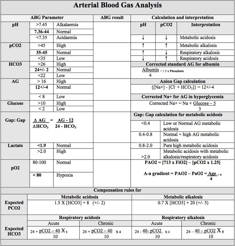
Homeostasis depends on the management of acid by-products and correction of acid-base imbalances via respiration (O2 and CO2 exchange) and various buffering systems. Illness can impact measurable items in the blood, by which their measurements (results) can be helpful in determining the severity of such illness.
Arterial Blood Gas Testing (ABGs)
An arterial blood gas (ABG) is the traditional method of estimating the:
- systemic carbon dioxide tension (PCO2) and
- pH, usually for the purpose of assessing ventilation and/or acid-base status.
It also measures:
- partial pressure of oxygen (PaO2--normal = 75-100 mmHg.),
- bicarbonate levels (HCO3--normal = 22-28 mEq/L), and
- oxygen saturation (SaO2--normal = 94-100%).
The value of arterial over venous sampling is that the success of oxygenation at the pulmonary level can be assessed, whereas venous gases are what's left from metabolism and have less relevance to the status of ventilation, respiration, oxygenation, or the elimination of CO2. In fact, venous oxygen (PvO2) has no practical value. Venous blood gasses can be used in a pinch, but the arterial version is simply superior and more informative and reliable.
The ABG measurements above are used for:
- Identification of an acid-base disturbance and monitoring its progression, via pH, bicarbonate (HCO3), and anion gap.
- Measurement of the partial pressures of oxygen (PaO2) and carbon dioxide (PaCO2).
- Assessing the patient's response to therapeutic interventions (e.g., insulin in patients with diabetic ketoacidosis).
ABGs are also useful in detecting abnormal hemoglobins (e.g., carbon monoxide poisoning) and for providing a blood sample that makes more tests available compared to a venous sample.
Note: pulse oximetry may be misleading in carbon monoxide poisoning, since carboxyhemoglobin absorbs at approximately the same frequency of 660 nm light as oxyhemoglobin, and this may be read as oxygen saturation. ABGs can be useful to eliminate the confusion.
In the field, there are special carbon monoxide oximeters that can detect the percentage of CO in the blood and differentiate it from CO2 and Oxygen.
ON THE ABGs REPORT:
- PaCO2 is the partial pressure of carbon dioxide. We can measure it to see how much respiratory acid (CO2) there is in the blood. (The a stands for "arterial'"; for the PCO2 of venous blood we would use PvCO2.)
The normal value for PaCO2 is 35-45 mmHg (mmHg is a means of identifying the pressure).
If CO2 is HIGH, it means there is a buildup of respiratory acids because the patient is not breathing enough CO2 away. Alternatively, it could mean that there is an excess build-up of CO2 from abnormal metabolism that compensatory breathing is not addressing.
- Bicarbonate
HCO3 measures the amount of bicarbonate in the bloodstream. HCO3 is the chemical name for bicarbonate, meaning the molecule is made up of one atom of hydrogen, one atom of carbon, and 3 atoms of oxygen.
The normal HCO3 level is 22-28/mEq/L (milliequivalents per liter.) This measures how much bicarbonate is in your blood binding up the excess acid.
If your HCO3 level is 10, it means that there is not enough of it, and so the acid in your blood just builds up and the overall pH of your body goes DOWN. This is called Metabolic Acidosis. To fix it, bring bicarbonate levels up by administering Bicarb via IV. If you overshoot, the excess bicarb binds up too much acid, so now the pH goes alkaline. This is called Metabolic Alkalosis. The ideal it to keep your patient stable long enough for the bicarb to bring down the levels of respiratory acid (CO2.)
Respiratory Acid-Base Abnormalities
In respiratory acidosis and alkalosis, it is the pulmonary system (lungs) and the resulting effects on CO2 that create the abnormality:
- If the pH is acidic (low-- <7 .35) and the CO2 is HIGH (>45), its considered Respiratory Acidosis (acid build up by retained CO2).
Causes: Hypoventilation (CNS depression), airway obstruction, pneumonia, pulmonary edema, pneumothorax, COPD, and sleep apnea.
- If pH is alkaline (high-- >7.45) and CO2 is LOW (<35), it means there are not enough respiratory acids because the patient is probably hyperventilating too much CO2 away. This is called Respiratory Alkalosis.
Causes: Hyperventilation, hypoxia, anxiety, sepsis.
Metabolic Acid-Base Abnormalities
In metabolic acidosis and alkalosis, cellular and organ injury create the abnormality, with a secondary influence by the lungs in an attempt to compensate:
- If the pH is acidic (low-- <7.35) and the CO2 is low (<35), this is Metabolic Acidosis. The low CO2 is a failed respiratory attempt at compensation by blowing off the respiratory acid (CO2). In metabolic acidosis, this rescue is simply not enough.
Causes: Uremia, alcohol, rhabdomyolisis.
- If the pH is alkaline (high-- >7.45) and the CO2 is high (>45), this is Metabolic Alkalosis.
Causes: Nausea and vomiting, alkali excess (antacids), diuretics.
In the Field
Field Note: On the trucks, pETCO2 (End Tidal Carbon Dioxide) is sometimes used and is expressed as a percentage. Instead of measuring the pressure in mmHg to measure the effectiveness of ventilations and circulation. A pETCO2 of 5-6% roughly equates to a PaCO2 of 35-45mmHg, so titrate your ventilations to achieve 5-6% pETCo2.
In the field, your most likely adventures into the world of acid-base abnormalities will be dealing with hypoxia, hypercapnea, sepsis, hypovolemia, and shock, where your main direction will be centered on support for rapid transport. This includes IV access, ABC maintenance (airway, breathing, and circulation).
Re: oxygen, It's hard to go wrong in administering oxygen for the purposes of assisting the lungs in its attempts to compensate for acidosis.
Not so fast! You actually CAN go wrong if the person has chronic hypercapnea (is immune to CO2 buildup from COPD, emphysema, or chronic interstitial lung disease). In these cases, of the two stimuli driving ventilation (high CO2 and low O2), only the low O2 stimulus is working:
GIVING SUCH A PERSON O2 WILL CAUSE RESPIRATORY ARREST because you acted on his or her low-O2 sensor that was all that was left keeping the breathing stimulus going.
How do you tell the difference?
There are varying degrees of abnormalities in gas exchange. Emphysema patients are called "pink puffers," because their PaO2 is only slightly reduced (they're still pink), but as COPD progresses and PaO2 is markedly reduced and PaCO2 is increased, they become "blue bloaters" (their color is dark). Any administration of O2 to a blue bloater is fraught with the possibility of respiratory failure if the patient is left to breathe on his or her own. Therefore, if O2 is given in these patients, it must be delivered by your actively (mechanically) ventilating the patient with an Ambu bag until a ventilator can continue to drive respiration artificially .